Journal of Clinical and Basic Research (JCBR)
Back to the articles list |
Back to browse issues page



1- Faculty of Medicine, Golestan University of Medical Sciences, Gorgan, Iran
2- Neuroscience Research Center, Biomedical Research Institute, Golestan University of Medical Sciences, Gorgan, Iran
3- Department Biochemistry and Medical Physics, Faculty of Medicine, Golestan University of Medical Sciences, Gorgan, Iran ,golshad.niknam@gmail.com
2- Neuroscience Research Center, Biomedical Research Institute, Golestan University of Medical Sciences, Gorgan, Iran
3- Department Biochemistry and Medical Physics, Faculty of Medicine, Golestan University of Medical Sciences, Gorgan, Iran ,
Full-Text [PDF 364 kb]
(333 Downloads)
| Abstract (HTML) (608 Views)
Discussion
The purpose of this study was to investigate the effects of RT on taste and smell perception in patients with HNC. This study was conducted on 25 Iranian patients. The cancer types examined included oral cavity cancer (50%), pharyngeal cancer (30%), and laryngeal cancer (20%). The results of the present study demonstrate that changes in taste and smell perception are side effects of RT in patients with HNC. Specifically, 65% of patients experienced changes in taste sensation, and 55% experienced changes in smell sensation. The most common taste change reported was a metallic taste, observed in 45% of patients. This side effect may be due to oxidative stress caused by RT and changes in saliva composition, which is consistent with findings from previous studies (17,18). Regarding smell sensation, a decreased ability to detect food odors (40%) was reported as the most frequent change, likely related to damage to the olfactory epithelium caused by the received radiation doses. Xerostomia was observed in 70% of patients and mucositis in 65%, both of which were identified as key factors exacerbating changes in taste and smell sensations. Xerostomia disrupts taste recognition due to reduced saliva production, while mucositis affects patients' sensory experience by causing inflammation in the mucosa of the mouth and throat. Data analysis showed that patients with oral cavity cancer experienced more taste changes. In contrast, patients with nasopharyngeal cancer reported more smell changes. Regarding demographic characteristics, older patients reported more sensory changes, which may be associated with age-related changes in sensory abilities or tissue sensitivity to radiation. Additionally, women reported more smell changes than men, which may be due to physiological differences or higher sensitivity to sensory changes. The findings of this study require further investigation in the future to clarify the underlying factors contributing to these differences. The obtained results may assist physicians in selecting medications during RT and help prevent harm to patients. They may also contribute to improving individuals' quality of life. Furthermore, reduced olfactory sensation in these patients may affect cognitive functions and various regions of the brain. All of these factors can impact the lives of these individuals.
In an article by Uchida et al., the relationship between olfactory dysfunction and cognitive ability, particularly attention, was investigated in the elderly population. The study demonstrated that a significant percentage of elderly participants exhibited olfactory dysfunction, and there was a notable correlation between impaired olfactory function and decreased cognitive performance. These findings suggest that olfactory dysfunction may serve as an early sign of cognitive decline in older adults (19). A systematic review and meta-analysis published by Yahya et al. examined the relationship between cognitive decline and structural and functional brain changes, as assessed by MRI, in patients with head and neck cancer following RT. The study found a significant correlation between cognitive performance, measured by the Montreal Cognitive Assessment (MoCA), and MRI changes in various brain regions, particularly the temporal lobe, which exhibited dose-dependent alterations (20). In addition, a study published by Álvarez-Camacho et al. examined olfactory disorders following RT for HNC. The results indicated that odor detection, identification, and discrimination are olfactory functions impaired after RT (21). A study published by Veyseller et al. investigated the impact of RT on olfactory bulb volume and olfactory function in patients with nasopharyngeal cancer. The findings indicated that RT leads to a reduction in olfactory bulb volume and a decline in olfactory function (22).
Despite providing valuable data, the present study had several limitations. First, the exclusion of patients who received concurrent chemotherapy limited the ability to examine the combined effects of these two treatment methods on taste and smell sensations. Second, the lack of longitudinal data to assess the improvement or persistence of sensory changes was another limitation. Third, although both subjective and objective tools were used, the standardization of these tools in the Iranian context may pose challenges, especially considering cultural differences and dietary habits. One key finding of this study was the significant association between severe sensory changes and reduced quality of life in patients. Decreased enjoyment of food, nutritional problems, and psychological impacts such as depression and social isolation were among the consequences of these changes.
Conclusion
The findings of this study confirm that changes in taste and smell are among the most common and impactful side effects of RT in patients with HNC. The prevalence of taste (65%) and smell alterations (55%), along with their association with xerostomia, mucositis, and diminished quality of life, highlights the need for strategies to manage these complications in clinical practice. Comparisons with other studies show relative consistency in the prevalence and types of sensory changes; however, variations in results may be related to treatment protocols, population characteristics, or the assessment tools used. The results of this study can help address issues affecting these patients and guide the selection of appropriate medications to prevent the resulting side effects.
Acknowledgement
The authors thank Golestan University of Medical Sciences and the Shafa Radiotherapy Center for their cooperation.
Funding Sources
Golestan University of Medical Sciences, Gorgan, Iran, provided financial support for this study with grant number.
Ethical Statement
This study was approved by the Ethics Committee of Golestan University of Medical Sciences with the approval code (Ethical code: IR.GOUMS.REC.1403.057).
Conflicts of Interest
The authors declare no conflicts of interest.
Author Contributions
This article is the result of a research project conducted in collaboration with Ms. Bahar Moslemi, Dr. Hamid Sepehri, and Alireza Khoshbin Khoshnazar at Golestan University of Medical Sciences.
Data Availability Statement
The data supporting the findings of this study are available from the corresponding author upon reasonable request.
Use of Artificial Intelligence
No artificial intelligence tools were used in the preparation of this assessment.
Full-Text: (69 Views)
Introduction
Head and neck cancer (HNC) remains one of the most common cancers and a significant cause of mortality (1). Radiotherapy (RT) is essential in the management of HNC, particularly in locally advanced cases. It can be used either as an independent definitive therapy or in conjunction with surgery, chemotherapy, or both. RT is a highly effective intervention that improves survival rates and may lead to cure (2). Radiation produces harmful free radicals within tissues, resulting in apoptosis and cell death, thereby eliminating rapidly dividing cancerous cells. However, radiation often affects malignant cells while also damaging adjacent healthy tissue (3-5). RT-related adverse effects are categorized according to their time of onset, ranging from immediate effects to late effects that may appear years after RT. The most prevalent acute effects arise from direct local irradiation-induced inflammatory responses, including painful mucositis, dysphagia, xerostomia, taste alterations, loss of appetite, and dermatitis. Late effects are primarily due to fibrosis in tissues that were previously inflamed (3,5,6).
Because healthy tissues are located close to the tumor bed, multiple side effects may frequently occur. These complications include dry mouth (Xerostomia), inflammation of the oral and pharyngeal mucosa (Mucositis), tissue fibrosis, osteoradionecrosis (Radiation-induced bone damage), and changes in taste and smell sensations (7). Such complications may be transient or persistent and can affect patients' quality of life, adherence to treatment, and clinical outcomes (8-11).
As reported in previous studies, a common side effect of these treatments is sensory impairment, which may involve deficits in smell and/or taste (12-14). These effects can negatively affect patients' quality of life and may contribute to malnutrition both during and after treatment.
The incidence of head and neck cancer has been increasing; nevertheless, advances in prevention, diagnosis, and treatment have contributed to a decline in mortality rates worldwide. Therefore, long-term outcomes and quality of life are becoming increasingly important for this patient population (12,15,16).
Considering that this concern can significantly affect patients' quality of life and may complicate or delay the treatment process, the aim of this study was to assess changes in olfactory and gustatory perception in patients undergoing RT at the Shafa Center, 5 Azar Hospital, Gorgan, in 2024, using a questionnaire.
Methods
Participants
The study involved a sample of 25 patients who received RT at the Shafa Center of 5 Azar Hospital in Gorgan, Iran (From March to January 2024). Patients were selected using a non-random (Convenience) sampling method and were recruited consecutively based on their visits to the hospital. This approach was adopted because of resource constraints, such as the number of patients and the RT schedule. In this study, an effort was made to maintain a male-to-female ratio of 3 to 1, consistent with epidemiological data on head and neck cancer (4). Patients aged between 18 and 70 were included to represent a broad age spectrum. The cancer types represented were oral cavity cancer (50%), pharyngeal cancer (30%), and laryngeal cancer (20%), indicating their relative prevalence in Iran (17).
Inclusion criteria
The inclusion criteria were defined as follows: a verified diagnosis of head and neck cancer established by TNM staging and pathological documents; an age range of 18 to 70 years to reduce the confounding effects of natural aging on sensory function; provision of informed consent by signing a consent form after a thorough and detailed explanation of the study objectives; sufficient cognitive ability to understand and respond to the questionnaires, as evaluated through clinical examination; and preserved olfactory and gustatory functions before the start of treatment.
Exclusion criteria
Exclusion criteria included the presence of sensory impairments before the initiation of RT (Including nerve damage or olfactory conditions); significant systemic illnesses (For instance, uncontrolled diabetes or Parkinson’s disease) known to influence sensory function; incomplete responses to questionnaires or withdrawal from the study; inability to communicate effectively (Such as in patients with serious speech disorders); and a previous history of chemotherapy. These criteria were established to ensure sample homogeneity and reduce confounding variables (7).
Experimental design
Scoring method
The data for this study were collected using questionnaires that assessed changes in olfactory and gustatory perceptions (Chemosensory Perception Questionnaire (CPQ)). The scoring method included 20 questions using a 5-point Likert scale, rated as follows: 0) no, 1) mild, 2) moderate, 3) severe, and 4) very severe. The questionnaire was divided into two sections: 10 questions related to olfaction and 10 questions related to gustation. Examples of gustatory questions were "Do you experience a metallic taste?" and "Has your ability to detect sweet tastes decreased?" Examples of olfactory questions were "Has your ability to detect the smell of food decreased?" and "Do you experience atypical odors?"
Implementation method
The questionnaire was completed with the help of the researcher and took approximately 15 to 20 minutes. Its reliability was evaluated using Cronbach’s alpha, which yielded a value of 0.85, indicating good internal consistency. The form was appropriate for individuals from different educational and cultural backgrounds.
Statistical analysis
The collected data were entered into SPSS statistical software version 20. Subsequently, the data distribution w:as char:acterized using descriptive statistics, including frequencies and percentages, as well as means and standard deviations.
Results
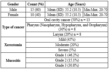
The results of this study are presented in the following tables: Table 1 shows the distribution of patients by demographic characteristics; Table 2 presents the prevalence and severity of changes in smell and taste sensations; and Table 3 displays the percentage of patients experiencing each type of taste change by cancer site.
Regarding treatment-related complications, mild xerostomia (Dry mouth) was the most prevalent (45%), while moderate and severe xerostomia were reported in 20% and 5% of patients, respectively. For mucositis, the majority of patients presented with Grade 1 (46.2%), followed by Grade 3 (30.8%) and Grade 2 (15.1%) (Table 1).
Among the 25 patients, taste alterations were more prevalent (65%) than olfactory changes (55%). Regarding taste sensation, the severity distribution was relatively balanced: moderate impairment was most common (25%), while mild and severe impairment each accounted for 20%. The most frequently reported taste changes were metallic taste (45%), followed by decreased sensitivity to sweet (35%), decreased sensitivity to salty (25%), and dysgeusia (15%). Regarding olfactory sensation, the prevalence was 55%, with severity distributed as follows: mild (15%), moderate (20%), and severe (20%). The most common olfactory changes included decreased recognition of food odors (40%), hyposmia (30%), and parosmia (20%) (Table 2).
Head and neck cancer (HNC) remains one of the most common cancers and a significant cause of mortality (1). Radiotherapy (RT) is essential in the management of HNC, particularly in locally advanced cases. It can be used either as an independent definitive therapy or in conjunction with surgery, chemotherapy, or both. RT is a highly effective intervention that improves survival rates and may lead to cure (2). Radiation produces harmful free radicals within tissues, resulting in apoptosis and cell death, thereby eliminating rapidly dividing cancerous cells. However, radiation often affects malignant cells while also damaging adjacent healthy tissue (3-5). RT-related adverse effects are categorized according to their time of onset, ranging from immediate effects to late effects that may appear years after RT. The most prevalent acute effects arise from direct local irradiation-induced inflammatory responses, including painful mucositis, dysphagia, xerostomia, taste alterations, loss of appetite, and dermatitis. Late effects are primarily due to fibrosis in tissues that were previously inflamed (3,5,6).
Because healthy tissues are located close to the tumor bed, multiple side effects may frequently occur. These complications include dry mouth (Xerostomia), inflammation of the oral and pharyngeal mucosa (Mucositis), tissue fibrosis, osteoradionecrosis (Radiation-induced bone damage), and changes in taste and smell sensations (7). Such complications may be transient or persistent and can affect patients' quality of life, adherence to treatment, and clinical outcomes (8-11).
As reported in previous studies, a common side effect of these treatments is sensory impairment, which may involve deficits in smell and/or taste (12-14). These effects can negatively affect patients' quality of life and may contribute to malnutrition both during and after treatment.
The incidence of head and neck cancer has been increasing; nevertheless, advances in prevention, diagnosis, and treatment have contributed to a decline in mortality rates worldwide. Therefore, long-term outcomes and quality of life are becoming increasingly important for this patient population (12,15,16).
Considering that this concern can significantly affect patients' quality of life and may complicate or delay the treatment process, the aim of this study was to assess changes in olfactory and gustatory perception in patients undergoing RT at the Shafa Center, 5 Azar Hospital, Gorgan, in 2024, using a questionnaire.
Methods
Participants
The study involved a sample of 25 patients who received RT at the Shafa Center of 5 Azar Hospital in Gorgan, Iran (From March to January 2024). Patients were selected using a non-random (Convenience) sampling method and were recruited consecutively based on their visits to the hospital. This approach was adopted because of resource constraints, such as the number of patients and the RT schedule. In this study, an effort was made to maintain a male-to-female ratio of 3 to 1, consistent with epidemiological data on head and neck cancer (4). Patients aged between 18 and 70 were included to represent a broad age spectrum. The cancer types represented were oral cavity cancer (50%), pharyngeal cancer (30%), and laryngeal cancer (20%), indicating their relative prevalence in Iran (17).
Inclusion criteria
The inclusion criteria were defined as follows: a verified diagnosis of head and neck cancer established by TNM staging and pathological documents; an age range of 18 to 70 years to reduce the confounding effects of natural aging on sensory function; provision of informed consent by signing a consent form after a thorough and detailed explanation of the study objectives; sufficient cognitive ability to understand and respond to the questionnaires, as evaluated through clinical examination; and preserved olfactory and gustatory functions before the start of treatment.
Exclusion criteria
Exclusion criteria included the presence of sensory impairments before the initiation of RT (Including nerve damage or olfactory conditions); significant systemic illnesses (For instance, uncontrolled diabetes or Parkinson’s disease) known to influence sensory function; incomplete responses to questionnaires or withdrawal from the study; inability to communicate effectively (Such as in patients with serious speech disorders); and a previous history of chemotherapy. These criteria were established to ensure sample homogeneity and reduce confounding variables (7).
Experimental design
Scoring method
The data for this study were collected using questionnaires that assessed changes in olfactory and gustatory perceptions (Chemosensory Perception Questionnaire (CPQ)). The scoring method included 20 questions using a 5-point Likert scale, rated as follows: 0) no, 1) mild, 2) moderate, 3) severe, and 4) very severe. The questionnaire was divided into two sections: 10 questions related to olfaction and 10 questions related to gustation. Examples of gustatory questions were "Do you experience a metallic taste?" and "Has your ability to detect sweet tastes decreased?" Examples of olfactory questions were "Has your ability to detect the smell of food decreased?" and "Do you experience atypical odors?"
Implementation method
The questionnaire was completed with the help of the researcher and took approximately 15 to 20 minutes. Its reliability was evaluated using Cronbach’s alpha, which yielded a value of 0.85, indicating good internal consistency. The form was appropriate for individuals from different educational and cultural backgrounds.
Statistical analysis
The collected data were entered into SPSS statistical software version 20. Subsequently, the data distribution w:as char:acterized using descriptive statistics, including frequencies and percentages, as well as means and standard deviations.
Results
The results of this study are presented in the following tables: Table 1 shows the distribution of patients by demographic characteristics; Table 2 presents the prevalence and severity of changes in smell and taste sensations; and Table 3 displays the percentage of patients experiencing each type of taste change by cancer site.
Regarding treatment-related complications, mild xerostomia (Dry mouth) was the most prevalent (45%), while moderate and severe xerostomia were reported in 20% and 5% of patients, respectively. For mucositis, the majority of patients presented with Grade 1 (46.2%), followed by Grade 3 (30.8%) and Grade 2 (15.1%) (Table 1).
Among the 25 patients, taste alterations were more prevalent (65%) than olfactory changes (55%). Regarding taste sensation, the severity distribution was relatively balanced: moderate impairment was most common (25%), while mild and severe impairment each accounted for 20%. The most frequently reported taste changes were metallic taste (45%), followed by decreased sensitivity to sweet (35%), decreased sensitivity to salty (25%), and dysgeusia (15%). Regarding olfactory sensation, the prevalence was 55%, with severity distributed as follows: mild (15%), moderate (20%), and severe (20%). The most common olfactory changes included decreased recognition of food odors (40%), hyposmia (30%), and parosmia (20%) (Table 2).

|
Table 3. Percentage of patients experiencing each type of taste change by cancer site
 |
Discussion
The purpose of this study was to investigate the effects of RT on taste and smell perception in patients with HNC. This study was conducted on 25 Iranian patients. The cancer types examined included oral cavity cancer (50%), pharyngeal cancer (30%), and laryngeal cancer (20%). The results of the present study demonstrate that changes in taste and smell perception are side effects of RT in patients with HNC. Specifically, 65% of patients experienced changes in taste sensation, and 55% experienced changes in smell sensation. The most common taste change reported was a metallic taste, observed in 45% of patients. This side effect may be due to oxidative stress caused by RT and changes in saliva composition, which is consistent with findings from previous studies (17,18). Regarding smell sensation, a decreased ability to detect food odors (40%) was reported as the most frequent change, likely related to damage to the olfactory epithelium caused by the received radiation doses. Xerostomia was observed in 70% of patients and mucositis in 65%, both of which were identified as key factors exacerbating changes in taste and smell sensations. Xerostomia disrupts taste recognition due to reduced saliva production, while mucositis affects patients' sensory experience by causing inflammation in the mucosa of the mouth and throat. Data analysis showed that patients with oral cavity cancer experienced more taste changes. In contrast, patients with nasopharyngeal cancer reported more smell changes. Regarding demographic characteristics, older patients reported more sensory changes, which may be associated with age-related changes in sensory abilities or tissue sensitivity to radiation. Additionally, women reported more smell changes than men, which may be due to physiological differences or higher sensitivity to sensory changes. The findings of this study require further investigation in the future to clarify the underlying factors contributing to these differences. The obtained results may assist physicians in selecting medications during RT and help prevent harm to patients. They may also contribute to improving individuals' quality of life. Furthermore, reduced olfactory sensation in these patients may affect cognitive functions and various regions of the brain. All of these factors can impact the lives of these individuals.
In an article by Uchida et al., the relationship between olfactory dysfunction and cognitive ability, particularly attention, was investigated in the elderly population. The study demonstrated that a significant percentage of elderly participants exhibited olfactory dysfunction, and there was a notable correlation between impaired olfactory function and decreased cognitive performance. These findings suggest that olfactory dysfunction may serve as an early sign of cognitive decline in older adults (19). A systematic review and meta-analysis published by Yahya et al. examined the relationship between cognitive decline and structural and functional brain changes, as assessed by MRI, in patients with head and neck cancer following RT. The study found a significant correlation between cognitive performance, measured by the Montreal Cognitive Assessment (MoCA), and MRI changes in various brain regions, particularly the temporal lobe, which exhibited dose-dependent alterations (20). In addition, a study published by Álvarez-Camacho et al. examined olfactory disorders following RT for HNC. The results indicated that odor detection, identification, and discrimination are olfactory functions impaired after RT (21). A study published by Veyseller et al. investigated the impact of RT on olfactory bulb volume and olfactory function in patients with nasopharyngeal cancer. The findings indicated that RT leads to a reduction in olfactory bulb volume and a decline in olfactory function (22).
Despite providing valuable data, the present study had several limitations. First, the exclusion of patients who received concurrent chemotherapy limited the ability to examine the combined effects of these two treatment methods on taste and smell sensations. Second, the lack of longitudinal data to assess the improvement or persistence of sensory changes was another limitation. Third, although both subjective and objective tools were used, the standardization of these tools in the Iranian context may pose challenges, especially considering cultural differences and dietary habits. One key finding of this study was the significant association between severe sensory changes and reduced quality of life in patients. Decreased enjoyment of food, nutritional problems, and psychological impacts such as depression and social isolation were among the consequences of these changes.
Conclusion
The findings of this study confirm that changes in taste and smell are among the most common and impactful side effects of RT in patients with HNC. The prevalence of taste (65%) and smell alterations (55%), along with their association with xerostomia, mucositis, and diminished quality of life, highlights the need for strategies to manage these complications in clinical practice. Comparisons with other studies show relative consistency in the prevalence and types of sensory changes; however, variations in results may be related to treatment protocols, population characteristics, or the assessment tools used. The results of this study can help address issues affecting these patients and guide the selection of appropriate medications to prevent the resulting side effects.
Acknowledgement
The authors thank Golestan University of Medical Sciences and the Shafa Radiotherapy Center for their cooperation.
Funding Sources
Golestan University of Medical Sciences, Gorgan, Iran, provided financial support for this study with grant number.
Ethical Statement
This study was approved by the Ethics Committee of Golestan University of Medical Sciences with the approval code (Ethical code: IR.GOUMS.REC.1403.057).
Conflicts of Interest
The authors declare no conflicts of interest.
Author Contributions
This article is the result of a research project conducted in collaboration with Ms. Bahar Moslemi, Dr. Hamid Sepehri, and Alireza Khoshbin Khoshnazar at Golestan University of Medical Sciences.
Data Availability Statement
The data supporting the findings of this study are available from the corresponding author upon reasonable request.
Use of Artificial Intelligence
No artificial intelligence tools were used in the preparation of this assessment.
References
1. Pandit PN, Patil RA, Palwe VS, Tambade SH, Wagh SY, Gavit MK, et al. Patterns of blood pressure changes in patients with head and neck cancer receiving radiotherapy or chemo-radiotherapy at a tertiary cancer centre. Head Neck. 2021;43(4):1280-8. [View at Publisher] [DOI] [PMID] [Google Scholar]
2. Leibowitz A, Grossman E, Berkovitch A, Levartovski M, Appel S, Sharabi Y, et al. The Effect of Head and Neck Radiotherapy on Blood Pressure and Orthostatic Hypotension in Patients With Head and Neck Tumors. Am J Hypertens. 2018;31(2):235-9. [View at Publisher] [DOI] [PMID] [Google Scholar]
3. Goyal M, Shukla P, Gupta D, Bisht S, Verma NS, Tiwari S, et al. Cardiovascular sequel of neck irradiation in head and neck cancer patients. Int J Radiat Biol. 2017;93(7):711-6. [View at Publisher] [DOI] [PMID] [Google Scholar]
4. Ferlay J, Soerjomataram I, Dikshit R, Eser S, Mathers C, Rebelo M, etr al. Cancer incidence and mortality worldwide: sources, methods and major patterns in GLOBOCAN 2012. Int J Cancer. 2015;136(5):E359-86. [View at Publisher] [DOI] [PMID] [Google Scholar]
5. Argiris A, Karamouzis MV, Raben D, Ferris RL. Head and neck cancer. Lancet. 2008;371(9625):1695-709. [View at Publisher] [DOI] [PMID] [Google Scholar]
6. Nutting CM, Morden JP, Harrington KJ, Urbano TG, Bhide SA, Clark C, et al. Parotid-sparing intensity modulated versus conventional radiotherapy in head and neck cancer (PARSPORT): a phase 3 multicentre randomised controlled trial. Lancet Oncol. 2011;12(2):127-36. [View at Publisher] [DOI] [PMID] [Google Scholar]
7. Vissink A, Mitchell JB, Baum BJ, Limesand KH, Jensen SB, Fox PC, et al. Clinical management of salivary gland hypofunction and xerostomia in head-and-neck cancer patients: successes and barriers. Int J Radiat Oncol Biol Phys. 2010;78(4):983-91. [View at Publisher] [DOI] [PMID] [Google Scholar]
8. Langendijk JA, Doornaert P, Verdonck-de Leeuw IM, Leemans CR, Aaronson NK, Slotman BJ. Impact of late treatment-related toxicity on quality of life among patients with head and neck cancer treated with radiotherapy. J Clin Oncol. 2008;26(22):3770-6. [View at Publisher] [DOI] [PMID] [Google Scholar]
9. Yamashita H, Nakagawa K, Tago M, Nakamura N, Shiraishi K, Eda M, et al. Taste dysfunction in patients receiving radiotherapy. Head Neck. 2006;28(6):508-16. [View at Publisher] [DOI] [PMID] [Google Scholar]
10. Ho W-K, Kwong DL, Wei WI, Sham JS. Change in olfaction after radiotherapy for nasopharyngeal cancer--a prospective study. Am J Otolaryngol. 2002;23(4):209-14. [View at Publisher] [DOI] [PMID] [Google Scholar]
11. Doty RL, Shah M, Bromley SM. Drug-induced taste disorders. Drug Saf. 2008;31(3):199-215. [View at Publisher] [DOI] [PMID] [Google Scholar]
12. Jha N, Speers J, Gastineau L, Patel S, Liu W, Pfahl E, et al. Smell and Taste Impairments in Head and Neck Cancer Patients-A Scoping Review. Nutrients. 2025;17(6):1087. [View at Publisher] [DOI] [PMID] [Google Scholar]
13. Schwartz LK, Weiffenbach JM, Valdez IH, Fox PC. Taste intensity performance in patients irradiated to the head and neck. Physiol Behav. 1993;53(4):671-7. [View at Publisher] [DOI] [PMID] [Google Scholar]
14. Jensen SB, Pedersen AML, Vissink A, Andersen E, Brown CG, Davies AN, et al . A systematic review of salivary gland hypofunction and xerostomia induced by cancer therapies: management strategies and economic impact. Support Care Cancer. 2010;18(8):1061-79. [View at Publisher] [DOI] [PMID] [Google Scholar]
15. Ravasco P, Monteiro-Grillo I, Marques Vidal P, Camilo ME. Impact of nutrition on outcome: a prospective randomized controlled trial in patients with head and neck cancer undergoing radiotherapy. Head Neck. 2005;27(8):659-68. [View at Publisher] [DOI] [PMID] [Google Scholar]
16. Epstein JB, Robertson M, Emerton S, Phillips N, Stevenson-Moore P. Quality of life and oral function in patients treated with radiation therapy for head and neck cancer. Head Neck. 2001;23(5):389-98. [View at Publisher] [DOI] [PMID] [Google Scholar]
17. Sharabi Y, Dendi R, Holmes C, Goldstein DS. Baroreflex failure as a late sequela of neck irradiation. Hypertension. 2003;42(1):110-6. [View at Publisher] [DOI] [PMID] [Google Scholar]
18. Wang X, Hu C, Eisbruch A. Organ-sparing radiation therapy for head and neck cancer. Nat Rev Clin Oncol. 2011;8(11):639-48. [View at Publisher] [DOI] [PMID] [Google Scholar]
19. Uchida S, Shimada C, Sakuma N, Kagitani F, Kan A, Awata S. The relationship between olfaction and cognitive function in the elderly. J Physiol Sci. 2020;70(1):48. [View at Publisher] [DOI] [PMID] [Google Scholar]
20. Voon NS, Abdul Manan H, Yahya N. Cognitive Decline following Radiotherapy of Head and Neck Cancer: Systematic Review and Meta-Analysis of MRI Correlates. Cancers (Basel). 2021;13(24):6191. [View at Publisher] [DOI] [PMID] [Google Scholar]
21. Álvarez-Camacho M, Gonella S, Campbell S, Scrimger RA, Wismer WV. A systematic review of smell alterations after radiotherapy for head and neck cancer. Cancer Treat Rev. 2017;54:110-21. [View at Publisher] [DOI] [PMID] [Google Scholar]
22. Veyseller B, Ozucer B, Degirmenci N, Gurbuz D, Tambas M, Altun M, et al. Olfactory bulb volume and olfactory function after radiotherapy in patients with nasopharyngeal cancer. Auris Nasus Larynx. 2014;41(5):436-40. [View at Publisher] [DOI] [PMID] [Google Scholar]
Send email to the article author
| Rights and permissions | |
 |
This work is licensed under a Creative Commons Attribution-NonCommercial 4.0 International License. |

This work is licensed under a Creative Commons Attribution-NonCommercial 4.0 International (CC BY-NC 4.0).
