Volume 9, Issue 1 (Journal of Clinical and Basic Research (JCBR) 2025)
jcbr 2025, 9(1): 20-23 |
Back to browse issues page


Download citation:
BibTeX | RIS | EndNote | Medlars | ProCite | Reference Manager | RefWorks
Send citation to:


BibTeX | RIS | EndNote | Medlars | ProCite | Reference Manager | RefWorks
Send citation to:
De Souza F M, Prabhu S P, Krishnan Dharan J. A cross-sectional morphometric study and clinical application of proximal end of humerus in Goan population. jcbr 2025; 9 (1) :20-23
URL: http://jcbr.goums.ac.ir/article-1-468-en.html
URL: http://jcbr.goums.ac.ir/article-1-468-en.html


1- Department of Anatomy, Goa Medical College, Bambolim, Goa, India
2- Department of Anatomy, Goa Medical College, Bambolim, Goa, India ,jai123krishnan@gmail.com
2- Department of Anatomy, Goa Medical College, Bambolim, Goa, India ,
Full-Text [PDF 573 kb]
(349 Downloads)
| Abstract (HTML) (1175 Views)
Results
The MHL was 309 ± 14.1 mm on the left side and 311 ± 16.5 mm on the right side, and the mean HHVD was 40.54 ± 3.1 mm on the left side and 40.38 ± 3.51 mm on the right side. Additionally, the mean HHTD was 37.84 ± 3.52 mm for the left humerus and 38.2 ± 3.8 mm for the right humerus.
The mean ANC was measured to be 126.9 ± 7.4 mm on the left side and 128.9 ± 11.2 mm on the right side. Moreover, the mean SNC was 83.2 ± 6.7 mm on the left side and 87.4 ± 9.7 mm on the right side.
The mean HHGT was measured as 10.64 ± 1.27 mm on the left side and 11.06 ± 0.98 mm on the right side. The mean BGW was 8.94 ± 1.64 mm on the left and 9.37 ± 1.6 mm on the right.
In our study, a statistically significant difference was observed between the right and left humeri with respect to the HHVD and the ANC, as detailed in Table 1.
Discussion
Acknowledging the substantial interspecimen and side-specific variability inherent in biological structures, dimensional variations constitute a critical factor that necessitates careful consideration during replacement surgery. Biomechanical studies have demonstrated that even minor deviations in prosthetic geometry from normative dimensions can compromise optimal functional outcomes.
The MHL observed in our study was 311 ± 16.5 mm for the right side and 309 ± 14.1 mm for the left side. These findings are consistent with those reported in previous studies by Prasad et al. (3), Rai et al. (4), and Jaiwal et al. (5).
The humeral head presents with a sphericity of slightly less than a hemisphere, a characteristic that, in comparison to the significantly smaller glenoid cavity of the scapula, predisposes the glenohumeral joint to instability and dislocation. Mechanical derangement of this articulation is a relatively common clinical entity, predominantly resulting from traumatic events. This is clinically significant due to the high propensity for recurrence following the initial dislocation. Glenohumeral dislocations can be classified as either anterior or posterior. Anterior dislocations are further subcategorized based on the final position of the humeral head relative to the glenoid fossa, namely preglenoid, subcoracoid, and subclavicular. Conversely, postero-inferior dislocations, wherein the humeral head resides in a subglenoid location, are observed with considerably less frequency (6).
To effectively perform Kocher's manoeuvre (Comprising positioning, external rotation, adduction, and internal rotation) for the reduction of shoulder dislocations, a comprehensive understanding of humeral head morphometry is crucial. This study involved the measurement of the HHTD and HHVD. The observed HHTD in the current research aligns with findings reported in previous studies by Prashant et al. (7), Jahan et al. (8), Kabakci et al. (9), and Sinha et al. (10).
The HHVD measured in our study was 40.38 ± 3.51 mm on the right side and 40.54 ± 3.1 mm on the left side. These findings contrast with those reported by Chatterjee et al. (11), who documented measurements of 35.52 ± 3.26 mm on the right and 35.19 ± 3.38 mm on the left. Furthermore, our results diverge from the observations of Ashutosh et al. (12), who found the HHVD to be 43.47 ± 3.92 mm on the right and 42.73 ± 3.55 mm on the left. In contrast to these discrepancies, the results obtained in the present study align with the findings of Prashant et al. (7) and Jahan et al. (8).
Our study revealed statistically significant disparities in the dimensions of the proximal humerus between the right and left sides within the studied cohort.
Furthermore, these findings diverged from previously reported anthropometric data for the Eastern Indian population. This discrepancy raises concerns regarding the applicability of current prosthetic designs, which often present relatively fixed dimensions despite some modularity, for achieving accurate anatomical restoration during shoulder arthroplasty, particularly in cases of traumatic injury (12).
The majority of injuries affecting the proximal humerus involve fractures localized at the surgical neck. These fractures exhibit a particularly high incidence in elderly individuals afflicted with osteoporosis, a condition characterized by diminished bone mineral density and consequent skeletal fragility. A fracture traversing the surgical neck of the humerus carries the potential to compromise the axillary nerve, which circumscribes this anatomical region. Furthermore, such fractures pose a risk of inducing avascular necrosis of the humeral head due to the potential disruption of the anastomotic vascular network situated around the surgical neck. Some patients necessitate surgical intervention, such as fracture fixation or arthroplasty, where a comprehensive understanding of the proximal humerus's dimensions is crucial. In this study, the mean SNC of the right humerus was measured at 83.2 ± 6.7 mm, while that of the left humerus was 87.4 ± 9.7 mm. These findings exhibit similarities to those reported by Kabakci et al. (9) in their investigation of a Turkish population.
The HHGT holds significant functional relevance in the abduction of the arm and clinical importance in the context of shoulder joint subluxation. Isolated fractures of the greater tubercle are frequently observed in adults and can, in some instances, exhibit substantial displacement (4) due to the tensile forces exerted by the supraspinatus muscle, which inserts onto this bony prominence. In cases of displaced fractures, open reduction and internal fixation may be indicated. Our research revealed a notable disparity in the HHGT measurements when compared to findings reported in previous studies by Jahan et al. (8), Rai et al. (4), Kabackci et al. (9), and Chatterjee et al. (11).
Full-Text: (300 Views)
Introduction
The humerus, the osseous structure of the arm and the longest bone of the superior limb, derives its nomenclature from the Latin term for the upper arm bone. Interestingly, the Greek word "omos" refers to the shoulder, highlighting the bone's proximal articulation. This long bone is characterized by distinct proximal and distal extremities, separated by a diaphysis. The proximal epiphysis comprises not only the humeral head, and the lesser and greater tubercles, but also the superior aspect of the intertubercular sulcus and the anatomical neck. Notably, the bicipital groove, or intertubercular sulcus, a groove situated between the greater and lesser tubercles, is located in the proximal third of the humerus. This groove serves as a conduit for the tendon of the long head of the biceps brachii muscle, its associated synovial sheath, and the ascending branch of the anterior circumflex humeral artery (1).
The glenohumeral joint exhibits the highest incidence of dislocation among all joints in the human body, with the majority of these events occurring in the anterior and inferior directions (2). Furthermore, the bicipital groove serves as a critical anatomical landmark for the accurate placement of shoulder prostheses. Pathologies affecting the long head of the biceps brachii have been implicated as a primary etiological factor in anterior shoulder pain, a prevalent musculoskeletal complaint within the Indian population. Traumatic impact to the shoulder commonly results in a non-displaced, comminuted fracture of the humeral greater tubercle in adult individuals, whereas elderly women are more susceptible to fractures through the surgical neck of the humerus. Post-surgical complications following fracture repair frequently include painful arc syndrome and adhesive capsulitis of the shoulder (2).
Therefore, comprehensive knowledge regarding the morphometry of the proximal humerus and the bicipital groove holds significant clinical and surgical relevance.
The objective of this study is to measure and analyze various morphometric parameters of the proximal humerus. Furthermore, this study aims to determine if statistically significant differences exist between these parameters when comparing the left and right humeri.
Methods
This observational study, conducted over a three-month period from November 2020 to January 2021, investigated 60 unpaired dry adult human humeri (31 left and 29 right) of undetermined age and gender. The specimens were obtained from the Department of Anatomy at Goa Medical College following ethical approval from the Institutional Ethics Committee. Epiphyseal closure was the criterion used to determine adulthood. Osteometric measurements were taken using an osteometric board, a digital vernier caliper with a precision of 0.01 mm, a measuring scale, and colored thread.
The following parameters were measured:
- Mean humeral length (MHL) - The distance between highest point of the humeral head and the lowest point of the trochlea;
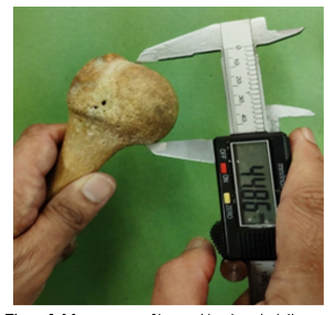
Humeral head vertical diameter (HHVD) - The diameter of the humeral head in the latero-medial direction (Figure 1);
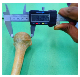
- Humeral head transverse diameter (HHTD) - The diameter of the humeral head in the antero-posterior direction (Figure 2);
- The distance between the highest point on the humeral head and the most proximal point of the greater tubercle (HHGT);
- Anatomical neck circumference (ANC);
- Surgical neck circumference (SNC);
- The distance from the lateral lip to the medial lip of the bicipital groove width (BGW)-measured between midpoint of medial and lateral lips (Figure 3).
Inclusion criteria
Dry adult human humeri of both genders were included in the study.
Exclusion criteria
Bones exhibiting macroscopic asymmetry or deformity, damaged osseous structures, and bones with a discernible epiphyseal line were excluded from the study.
Statistical analysis
The data underwent statistical analysis employing an independent samples t-test, conducted with SPSS software (Version 24).
.PNG)
Results
The MHL was 309 ± 14.1 mm on the left side and 311 ± 16.5 mm on the right side, and the mean HHVD was 40.54 ± 3.1 mm on the left side and 40.38 ± 3.51 mm on the right side. Additionally, the mean HHTD was 37.84 ± 3.52 mm for the left humerus and 38.2 ± 3.8 mm for the right humerus.
The mean ANC was measured to be 126.9 ± 7.4 mm on the left side and 128.9 ± 11.2 mm on the right side. Moreover, the mean SNC was 83.2 ± 6.7 mm on the left side and 87.4 ± 9.7 mm on the right side.
The mean HHGT was measured as 10.64 ± 1.27 mm on the left side and 11.06 ± 0.98 mm on the right side. The mean BGW was 8.94 ± 1.64 mm on the left and 9.37 ± 1.6 mm on the right.
In our study, a statistically significant difference was observed between the right and left humeri with respect to the HHVD and the ANC, as detailed in Table 1.
Table 1. Measurements of proximal end of the humerus in our study.PNG) |
Discussion
Acknowledging the substantial interspecimen and side-specific variability inherent in biological structures, dimensional variations constitute a critical factor that necessitates careful consideration during replacement surgery. Biomechanical studies have demonstrated that even minor deviations in prosthetic geometry from normative dimensions can compromise optimal functional outcomes.
The MHL observed in our study was 311 ± 16.5 mm for the right side and 309 ± 14.1 mm for the left side. These findings are consistent with those reported in previous studies by Prasad et al. (3), Rai et al. (4), and Jaiwal et al. (5).
The humeral head presents with a sphericity of slightly less than a hemisphere, a characteristic that, in comparison to the significantly smaller glenoid cavity of the scapula, predisposes the glenohumeral joint to instability and dislocation. Mechanical derangement of this articulation is a relatively common clinical entity, predominantly resulting from traumatic events. This is clinically significant due to the high propensity for recurrence following the initial dislocation. Glenohumeral dislocations can be classified as either anterior or posterior. Anterior dislocations are further subcategorized based on the final position of the humeral head relative to the glenoid fossa, namely preglenoid, subcoracoid, and subclavicular. Conversely, postero-inferior dislocations, wherein the humeral head resides in a subglenoid location, are observed with considerably less frequency (6).
To effectively perform Kocher's manoeuvre (Comprising positioning, external rotation, adduction, and internal rotation) for the reduction of shoulder dislocations, a comprehensive understanding of humeral head morphometry is crucial. This study involved the measurement of the HHTD and HHVD. The observed HHTD in the current research aligns with findings reported in previous studies by Prashant et al. (7), Jahan et al. (8), Kabakci et al. (9), and Sinha et al. (10).
The HHVD measured in our study was 40.38 ± 3.51 mm on the right side and 40.54 ± 3.1 mm on the left side. These findings contrast with those reported by Chatterjee et al. (11), who documented measurements of 35.52 ± 3.26 mm on the right and 35.19 ± 3.38 mm on the left. Furthermore, our results diverge from the observations of Ashutosh et al. (12), who found the HHVD to be 43.47 ± 3.92 mm on the right and 42.73 ± 3.55 mm on the left. In contrast to these discrepancies, the results obtained in the present study align with the findings of Prashant et al. (7) and Jahan et al. (8).
Our study revealed statistically significant disparities in the dimensions of the proximal humerus between the right and left sides within the studied cohort.
Furthermore, these findings diverged from previously reported anthropometric data for the Eastern Indian population. This discrepancy raises concerns regarding the applicability of current prosthetic designs, which often present relatively fixed dimensions despite some modularity, for achieving accurate anatomical restoration during shoulder arthroplasty, particularly in cases of traumatic injury (12).
The majority of injuries affecting the proximal humerus involve fractures localized at the surgical neck. These fractures exhibit a particularly high incidence in elderly individuals afflicted with osteoporosis, a condition characterized by diminished bone mineral density and consequent skeletal fragility. A fracture traversing the surgical neck of the humerus carries the potential to compromise the axillary nerve, which circumscribes this anatomical region. Furthermore, such fractures pose a risk of inducing avascular necrosis of the humeral head due to the potential disruption of the anastomotic vascular network situated around the surgical neck. Some patients necessitate surgical intervention, such as fracture fixation or arthroplasty, where a comprehensive understanding of the proximal humerus's dimensions is crucial. In this study, the mean SNC of the right humerus was measured at 83.2 ± 6.7 mm, while that of the left humerus was 87.4 ± 9.7 mm. These findings exhibit similarities to those reported by Kabakci et al. (9) in their investigation of a Turkish population.
The HHGT holds significant functional relevance in the abduction of the arm and clinical importance in the context of shoulder joint subluxation. Isolated fractures of the greater tubercle are frequently observed in adults and can, in some instances, exhibit substantial displacement (4) due to the tensile forces exerted by the supraspinatus muscle, which inserts onto this bony prominence. In cases of displaced fractures, open reduction and internal fixation may be indicated. Our research revealed a notable disparity in the HHGT measurements when compared to findings reported in previous studies by Jahan et al. (8), Rai et al. (4), Kabackci et al. (9), and Chatterjee et al. (11).
|
Table 2. Comparing the measurements from our study and those from other studies
.PNG) |
An isolated fracture involving the anatomical neck of the humerus represents an exceedingly uncommon pathological occurrence within the domain of orthopaedic surgery. Avascular necrosis stands as one of the most significant and concerning complications associated with this specific fracture pattern.
Pathologies affecting the tendon of the long head of the biceps brachii are a primary etiology of anterior shoulder pain. Given the close anatomical relationship between the long head of the biceps tendon and the bicipital groove, the morphometry of the latter is a significant consideration. The bicipital groove serves as a crucial anatomical landmark for the placement of the lateral fin of shoulder joint prostheses; therefore, its morphometric characteristics are pertinent to the design, positioning, and sizing of these prosthetic devices. BGW is a clinically significant anatomical consideration, as an increased width is implicated in the potential for subluxation of the biceps tendon. In such cases of instability, a recommended surgical intervention involves securing the tendon to the floor of the bicipital groove, coupled with the resection of the superior portion of the groove (13). Narrow bicipital groove can culminate in impingement of the long head of the biceps brachii tendon, resulting in anterior shoulder pain. The biceps brachii muscle is actively involved in strenuous physical labor and often exhibits hypertrophy in individuals engaged in manual occupations. The increased mechanical stress exerted on the bicipital groove by a hypertrophied biceps tendon can consequently deepen and widen this groove. Given the significant influence of occupation on the structural characteristics of the bicipital groove, regional variations in its morphometry are anticipated. Therefore, a morphometric analysis of BGW is clinically relevant, as it provides surgeons with crucial data regarding BGW within a specific population. Our research on BGW yielded findings consistent with Kabakci et al.’s (9) study, yet diverged from the outcomes reported by Rajan et al. (14).
Table 2 presents a comparison between the measurements from our study and those reported in the aforementioned articles.
The demographic characteristics of the studied cohort, including age, occupation, and nutritional status, were not ascertained in this research. The current study centered on the morphometric analysis of the proximal humerus bilaterally; however, it did not explore variations based on gender. Given the documented functional disparities in the shoulder joint between biological genders, potential gender-related differences in humeral morphology may exist and warrant further investigation to elucidate their nature and extent.
Conclusion
This study reveals notable racial and ethnic variations in specific anthropometric measurements, potentially enhancing the understanding of the proximal humerus's morphology and functional characteristics. These findings could be valuable for optimizing the planning of orthopedic surgical interventions targeting the proximal humerus.
This research holds the potential to inform prosthesis manufacturers in the design and customization of their products, taking into account ethnic and racial variations.
The morphometric characteristics of the bicipital groove are clinically significant, as a shallow groove is associated with an elevated risk of biceps tendon subluxation.
Furthermore, the findings of this study may prove beneficial for radiologists, anthropologists, and forensic experts in their respective professional practices.
Acknowledgement
We would like to thank Dr. Manoj Kumar Kulkarni, statistician and associate professor in the Department of Community Medicine at Goa Medical College, for his assistance in conducting the statistical analysis.
Funding sources
None.
Ethical statement
This study received approval from the Institutional Ethics Committee of Goa Medical College, as documented in letter No. GMC/IEC/Dec-20/02.
Conflicts of interest
None.
Author contributions
FMDS: Conceptualization, supervision, validation, and review and editing of the draft; SPP: Data collection, measurement and analysis, and review and editing of the draft; JKD: Data collection, measurement and analysis, visualisation of data, and preparing the initial draft.
Data availability statement
Data can be provided upon request.
Article Type: Research |
Subject:
Basic medical sciences
References
1. Drake Richard, Vogl W, Mitchelle Adam, Tibbitts R, Richardson P, Gray H. Gray's Anatomy for Students. 3rd ed. Philadelphia:Churchill Livingstone Elsevier;2015. p.704 [View at Publisher] [Google Scholar]
2. Maheshwari J, Mhaskar VA. Essential orthopaedics. 5th ed. Delhi:Jaypee brothers;2015. p.93 [View at Publisher]
3. Prasad NC, Shivashankarappa A, Pavan PH, Shruthi BN, Saheb SH. A study on segments of humerus and its clinical importance. International Journal of Orthopaedics Sciences. 2017;3(4k):752-4. [View at Publisher] [DOI] [Google Scholar]
4. Rai R, Chawla M. Morphometry of adult humerus bone in Moradabad region. IJBAR. 2014;05(03):163-5. [View at Publisher] [DOI] [Google Scholar]
5. Jaiswal P, Kumar Verma R. Analysis of morphometric segments of humerus with clinical relevance in Rajasthan region. Int J Med Sci Educ. 2019;6(2):121-7. [View at Publisher] [Google Scholar]
6. Moore KL, Dalley AF, Agur AMR. Clinically oriented anatomy . 8th ed. Philadelphia:Wolters Kluwer;2018. p.815 [View at Publisher] [Google Scholar]
7. Prashant KU, Pai MM, Murlimanju BV, Prabhu LV, Prameela MD. Estimation of the humerus length by its proximal segments: A South Indian anatomical study. J Morphol Sci. 2019;36(02):067-71. [View at Publisher] [DOI] [Google Scholar]
8. Jahan S, Srivastava R. Morphometric study of proximal end of humerus in North Indian population. JMSCR . 2020 Aug;08(08):102-6. [View at Publisher] [DOI] [Google Scholar]
9. Kabakci A, Buyukmumcu M, Yilmaz MT, Cicekcibasi AE, Akın D, Cihan E. An Osteometric Study on Humerus. Int J Morphol. 2017;35(1):219-26. [View at Publisher] [DOI] [Google Scholar]
10. Sinha P, LakhiBhutia K, Tamang KB. Morphometric measurements of segments in dry humerus. J Evol Med Dent Sci. 2017;6(67):4819-22. [View at Publisher] [DOI] [Google Scholar]
11. Chatterjee M, Sinha I, Poddar R, Ghosal AK. Humeral morphometrics: A study in Eastern Indian Population. IJAR. 2017;5(4.1):4454-69. [View at Publisher] [DOI] [Google Scholar]
12. Ashutosha A, Deepali RK, Ajay C, BH B, Ashish B. Morphometric analysis and surgical anatomy of proximal humerus. IJAR. 2017;5(3.1):4056-62. [View at Publisher] [DOI] [Google Scholar]
13. Rajani S, Man S. Review of bicipital groove morphology and its analysis in North Indian population. ISRN Anat. 2013;2013:243780. [View at Publisher] [DOI] [PMID] [Google Scholar]
14. Rajan YS, Sampath Kumar SK. Morphometric study on bicipital groove among South Indian population. JCDR. 2016;10(7):AC01-3. [View at Publisher] [DOI] [PMID] [Google Scholar]
Send email to the article author
| Rights and permissions | |
 |
This work is licensed under a Creative Commons Attribution-NonCommercial 4.0 International License. |

This work is licensed under a Creative Commons Attribution-NonCommercial 4.0 International (CC BY-NC 4.0).
